
Zwitterionic hydrogel coating for medical devices
The surface the body ignores.
ZwiGuard is a zwitterionic hydrogel coating, covalently bonded to the device, that keeps protein, cells and bacteria off. Less scar, lower impedance, easier insertion, fewer bacteria. One coating step. Ten years of evidence.
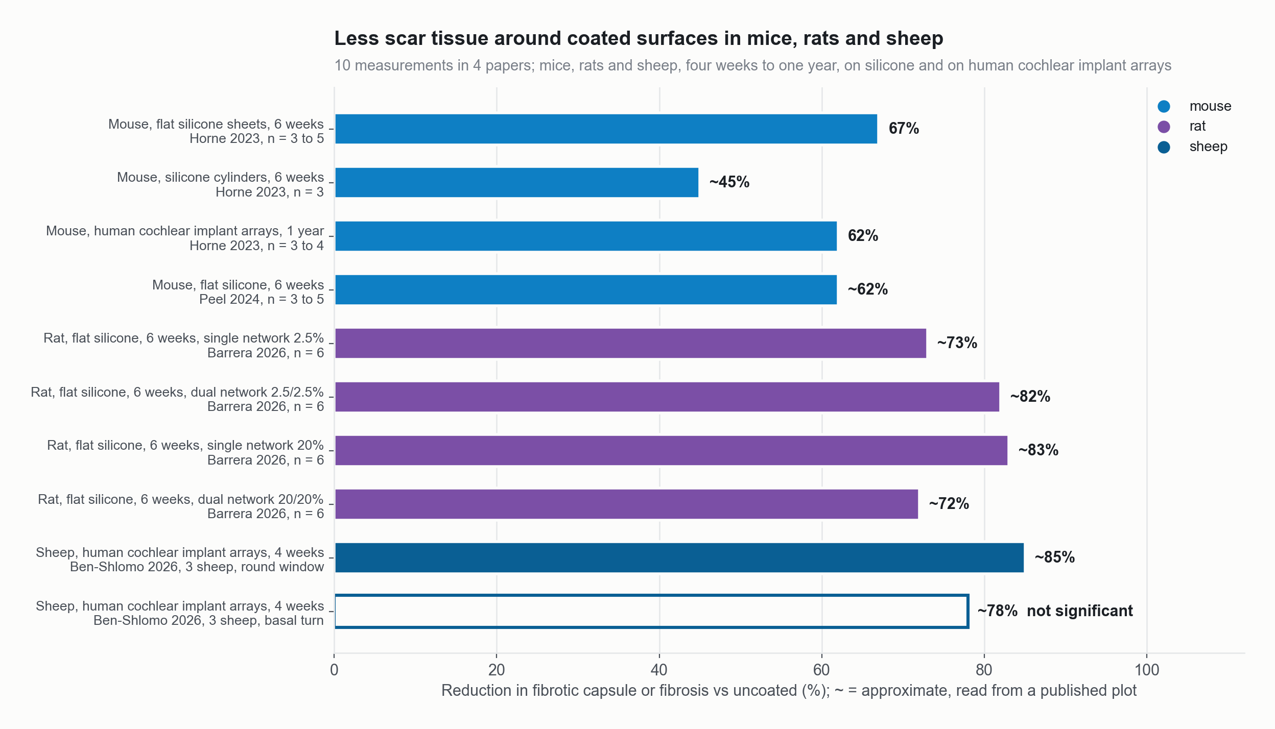
Less scar tissue around coated surfaces in mice, rats and sheep: percent reduction in fibrotic capsule or fibrosis, measurement by measurement. The hollow bar is not statistically significant.
Source: 10 measurements in mice, rats and sheep. Horne 2023, Acta Biomaterialia; Peel 2024, ACS Applied Bio Materials; Barrera 2026, ACS Applied Polymer Materials; Ben-Shlomo 2026, Hearing Research. Values read from the published figures; the basal-turn bar is not significant.
- 01 Thicknessa few micrometres to 100 or more, tunable
- 02 Bondgel tears before the interface lets go (Leigh 2019)
- 03 Impedanceabout half, four weeks in sheep (Ben-Shlomo 2026)
The public record in numbers
- 10peer-reviewed papers, 2017 to 2026Protein, cells, bacteria and scar all measured, in vitro and in four animal and cadaver models.
- 3PhD dissertations, University of IowaThree doctoral theses underpin the chemistry and the process.
- 3issued US patentsThe photografting method and the coated-device system are protected.
- 7patents granted in five countriesUnited States, Australia, China, Japan and Mexico: the largest device markets.
- 5provisional applications filedFive new inventions filed; national applications pending in Canada, Europe and South Africa.
- 1 yearfilm intact after implantation, miceDurable: the covalent graft outlasts the study and the capsule stays thin.
- 4 weeksfunctional human arrays in sheepWorks on the real device: lower impedance and less inflammation on a working implant.
- ~90%less protein, cell and bacterial adhesion and friction, published bench studiesNothing sticks: the hydration shell turns away protein, cells and bacteria before scar or biofilm can start.
Every number on this site comes from a published paper, dissertation or issued patent.
And behind the papers: 182 archived experiments, 1,056 adhesion tests, 136 animals.
Sources: the ten journal papers and the dissertations listed on the Publications page, USPTO records, ZCMI patent schedule, 2025.
Less scar
Scar tissue was about 45 to 85 percent lower around coated surfaces.
Ten measurements from four papers, in mice, rats and sheep, from four weeks to one year, on silicone and on real implant arrays. The sheep basal-turn measurement was not statistically significant.
In mice, the capsule was 67 percent thinner at six weeks on flat silicone and 62 percent thinner at one year on coated cochlear implant arrays (Horne 2023, Acta Biomaterialia).
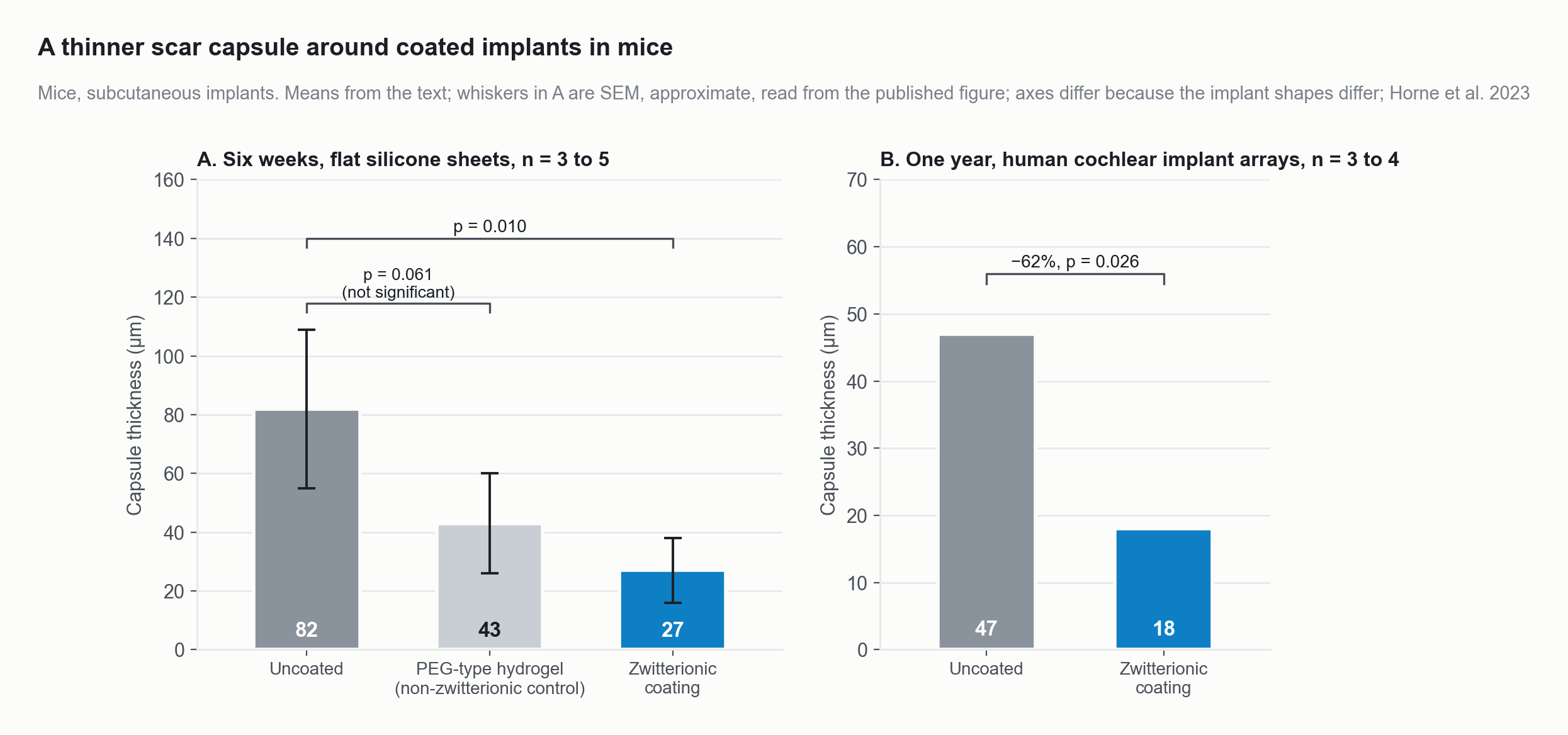
In mice, scar capsules were 67 percent thinner around zwitterionic-coated silicone at six weeks and 62 percent thinner around coated human cochlear implant arrays at one year. A non-zwitterionic hydrogel control did not differ significantly from uncoated.
Source: Horne R, Ben-Shlomo N, Jensen M, et al. Acta Biomaterialia 2023;166:212-223, Figs 5D and 7I (means and p values from the text; SEM whiskers approximate, read from the published figure). doi:10.1016/j.actbio.2023.05.011
Lower impedance
Coated implant arrays read about half the impedance of uncoated ones.
Working human cochlear implant arrays in sheep read 67 percent lower impedance at implantation and 47 percent lower four weeks later.
Three sheep, one coated and one uncoated array each (Ben-Shlomo 2026, Hearing Research). No coated device has been implanted in a person.
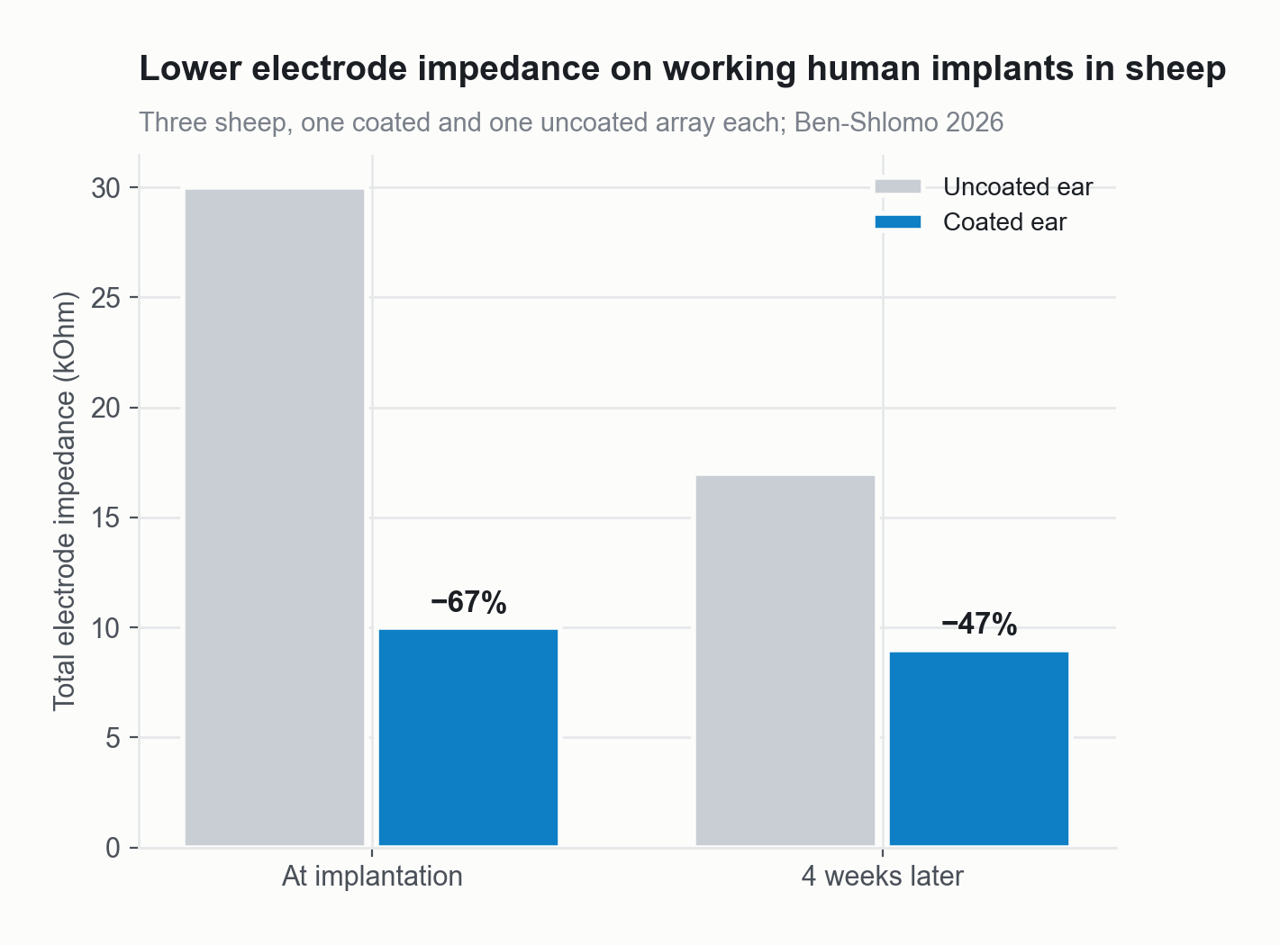
Electrode impedance of coated versus uncoated cochlear implant arrays in sheep.
Source: Ben-Shlomo et al. 2026, Hearing Research 474:109606, Fig 3B (values read from the figure).
Easier insertion
Insertion force fell by about 40 percent.
Coated cochlear implant arrays needed 37.0 mN of peak force against 64.2 mN for uncoated arrays, in human cadaveric cochleae (n = 9 per group, p = 0.001).
Force variation during insertion fell from 89.9 to 52.1 mN/s (p = 0.03). Bennion 2021, Otology and Neurotology.
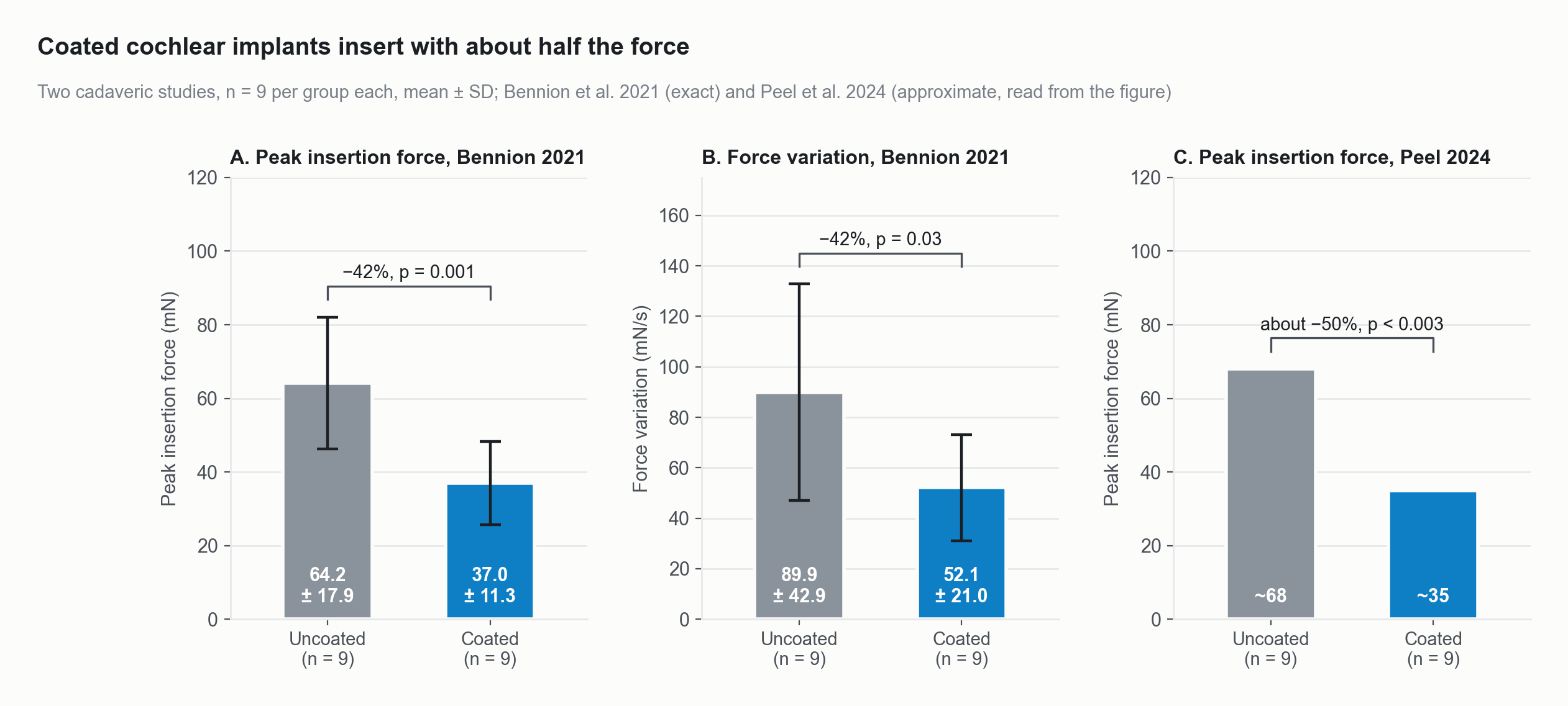
Coated cochlear implant arrays needed about 40 percent less peak force, and had about 42 percent less force variation, than uncoated arrays in human cadaveric cochleae.
Source: Bennion DM, Horne R, Peel A, Reineke P, Henslee A, Kaufmann C, Guymon CA, Hansen MR. Otology and Neurotology 2021;42(10):1476-1483. doi:10.1097/MAO.0000000000003288 (Fig 4A and 4B; means and SD from the text).
Fewer bacteria, less friction
Protein, cells, bacteria and friction all fall by about 90 percent.
Across 38 published comparisons of coated and uncoated surfaces, from eight papers, the median reduction was 94 percent. The range was 53 to 99 percent.
The four groups are fibrinogen adsorption, cell attachment, S. aureus and S. epidermidis adhesion, and friction. These are laboratory and explant measurements, not clinical results.
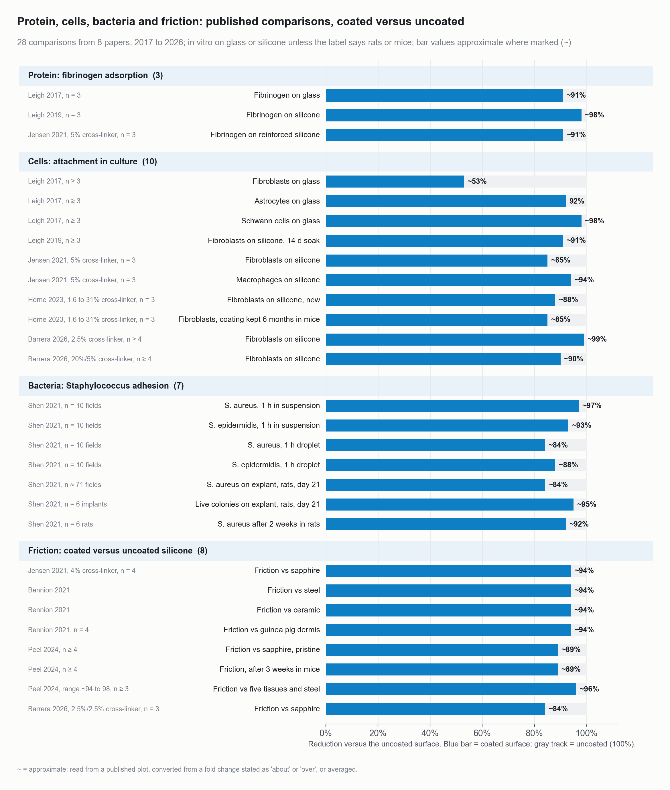
Protein, cells, bacteria and friction: every published bench comparison of coated versus uncoated surfaces (38 bars from 8 papers), as percent reduction.
Source: 38 comparisons from 8 papers, 2017 to 2026: Leigh 2017 Biomacromolecules; Leigh 2019 Langmuir; Jensen 2021 ACS Biomaterials Science and Engineering; Shen 2021 ACS Applied Bio Materials; Bennion 2021 Otology and Neurotology; Horne 2023 Acta Biomaterialia; Peel 2024 ACS Applied Bio Materials; Barrera 2026 ACS Applied Polymer Materials. Bar values approximate where marked, read from the published plots.
It stays on
The film was still intact at one year in mice.
Checkpoints run from two weeks in rats to one year in mice. Antifouling was unchanged at six months (Horne 2023, Peel 2024).
The hydrogel is covalently grafted, so it is part of the surface rather than a layer on top of it.
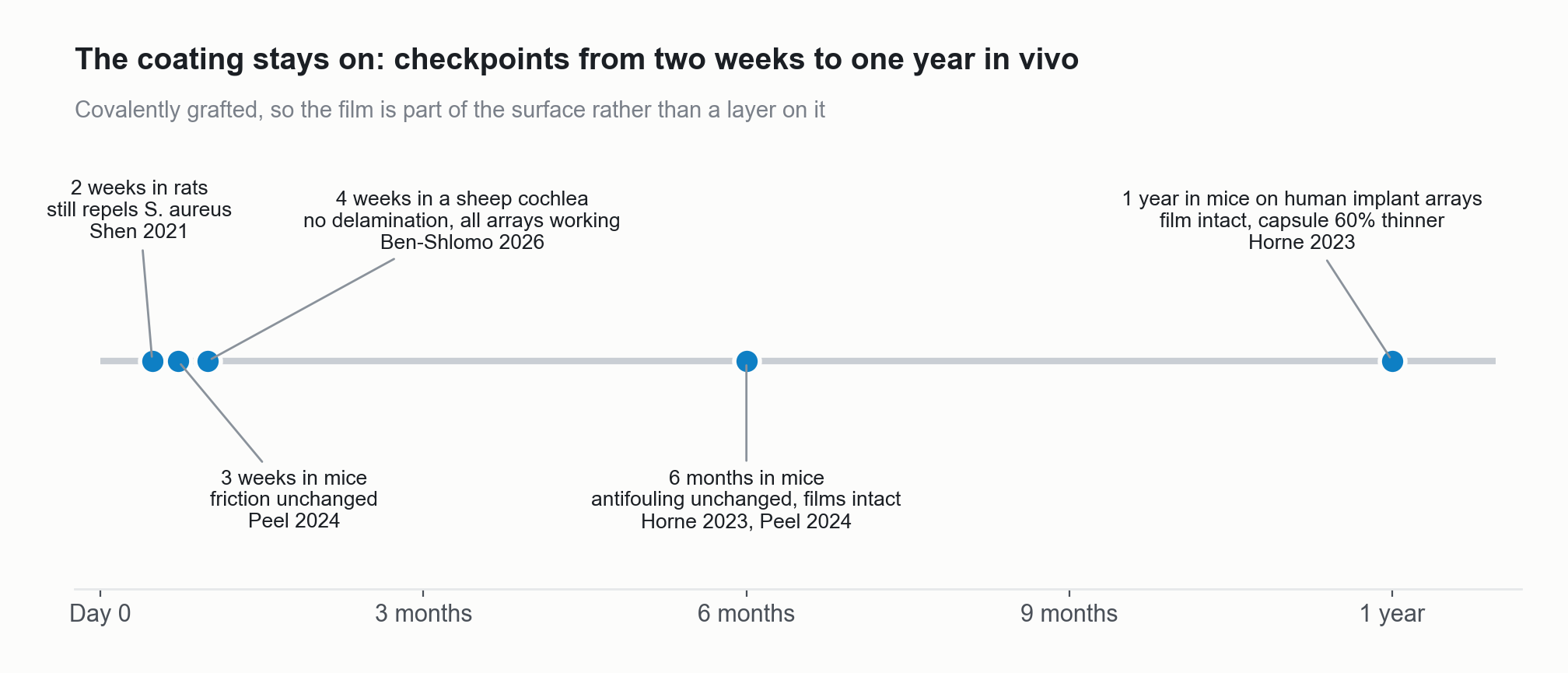
In-vivo durability checkpoints from two weeks to one year.
Source: Shen 2021; Peel 2024; Ben-Shlomo 2026; Horne 2023.
The process
One dip, one UV step.
The part is dipped into the monomer solution, then moved under UV light. One UV step grafts the film and crosslinks it.
Applications
Nine device families, one number that matters for each.
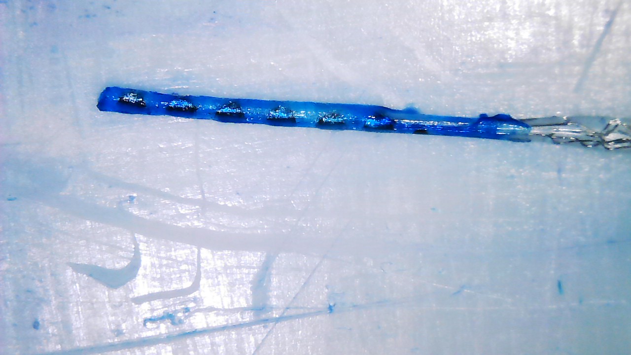
Neural interfaces
~50%
lower total electrode impedance after four weeks in sheep (Ben-Shlomo 2026)
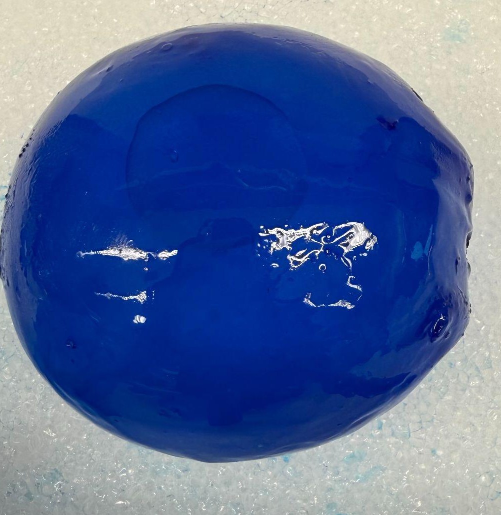
Tissue expanders and breast implants
82 to 27 µm
capsule thickness around coated silicone in mice at six weeks (Horne 2023)
Vascular access
~97%
less S. aureus adhesion on coated silicone in vitro (Shen 2021)
Indwelling catheters
12.5 vs 12.6%
symptomatic CAUTI with silver alloy versus standard catheters: the problem a new surface must solve (Pickard 2012)
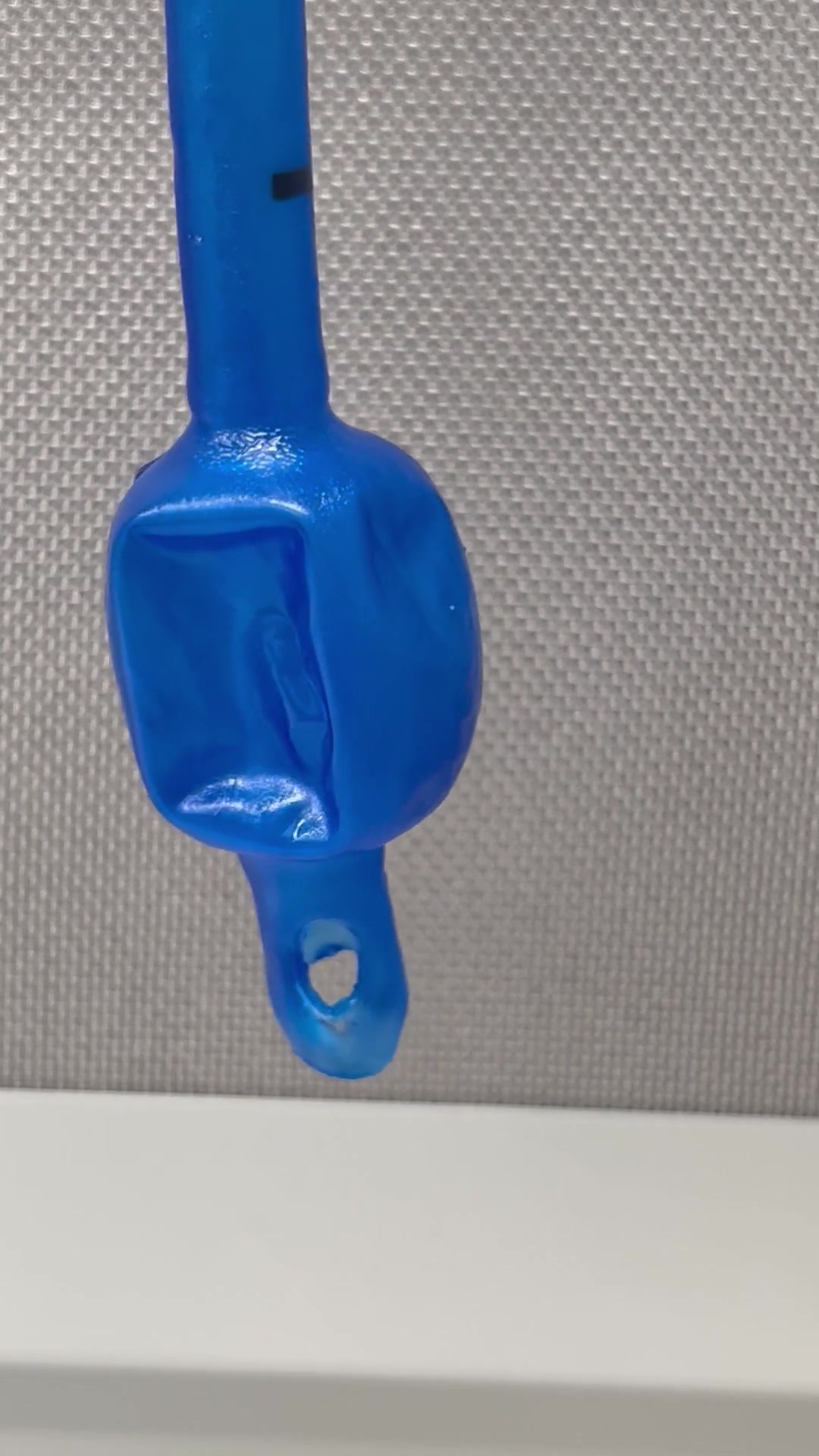
Airway tubes
~40%
less peak insertion force for coated cochlear implant arrays in human cadaveric cochleae (Bennion 2021)
GI stents
22.5 vs 30 min
median time to clear a dried mucus plug, coated against uncoated tubes (Horne 2025)
PEEK and orthopedic
feasibility
PEEK bonding is measured on your parts
Sensors
~60%
thinner capsule around implanted arrays at one year in mice (Horne 2023)
ENT and feeding tubes
~0°
tilt to start saline flow on coated silicone sheets, against 45 uncoated (Horne 2025)
How we work together
Three steps, and the first one is free.
Feasibility, free
You send parts. We coat them, with no cap on quantity, and return them with matched uncoated controls and our data.
Joint development
We optimize the coating on your production devices and run ISO 10993, sterilization, aging and an animal model.
Manufacturing
We coat for you. Our FDA Device Master File sits behind your submission.
- Origin
- University of Iowa spin-off.
- Funding
- NIH SBIR Phase I and Phase II.
- Patents
- US 11,525,069; 11,814,526; 12,116,498.
- Papers
- Ten peer-reviewed papers.
Bring us a device with a fouling, scar or friction problem.
We coat it free, with no cap on quantity, and return it with matched uncoated controls and data.