
Application
Sensors
ZwiGuard™ is a drug-free coating tested for scar capsule thickness on silicone and implant arrays, with sensor response still to be measured on your parts.
- Devices
- Continuous glucose monitors, implanted biosensors
- Substrates
- silicone and platinum contacts (published); PVC, PEEK, parylene-C and polyurethane (ZwiCoat internal tests); sensor membranes in the feasibility round
- Thickness
- tunable, from a few micrometres to 100 micrometres or more (ZwiCoat process range); about 7 micrometres in the published silicone work (Leigh 2019)
- Sterilization
- validated on your cycle during joint development
- Regulatory path
- 510(k) or PMA supplement with an accuracy study, set by your device; our Master File supports the filing
- Evidence
- mice, six weeks to one year (silicone and arrays); sensor response not yet measured
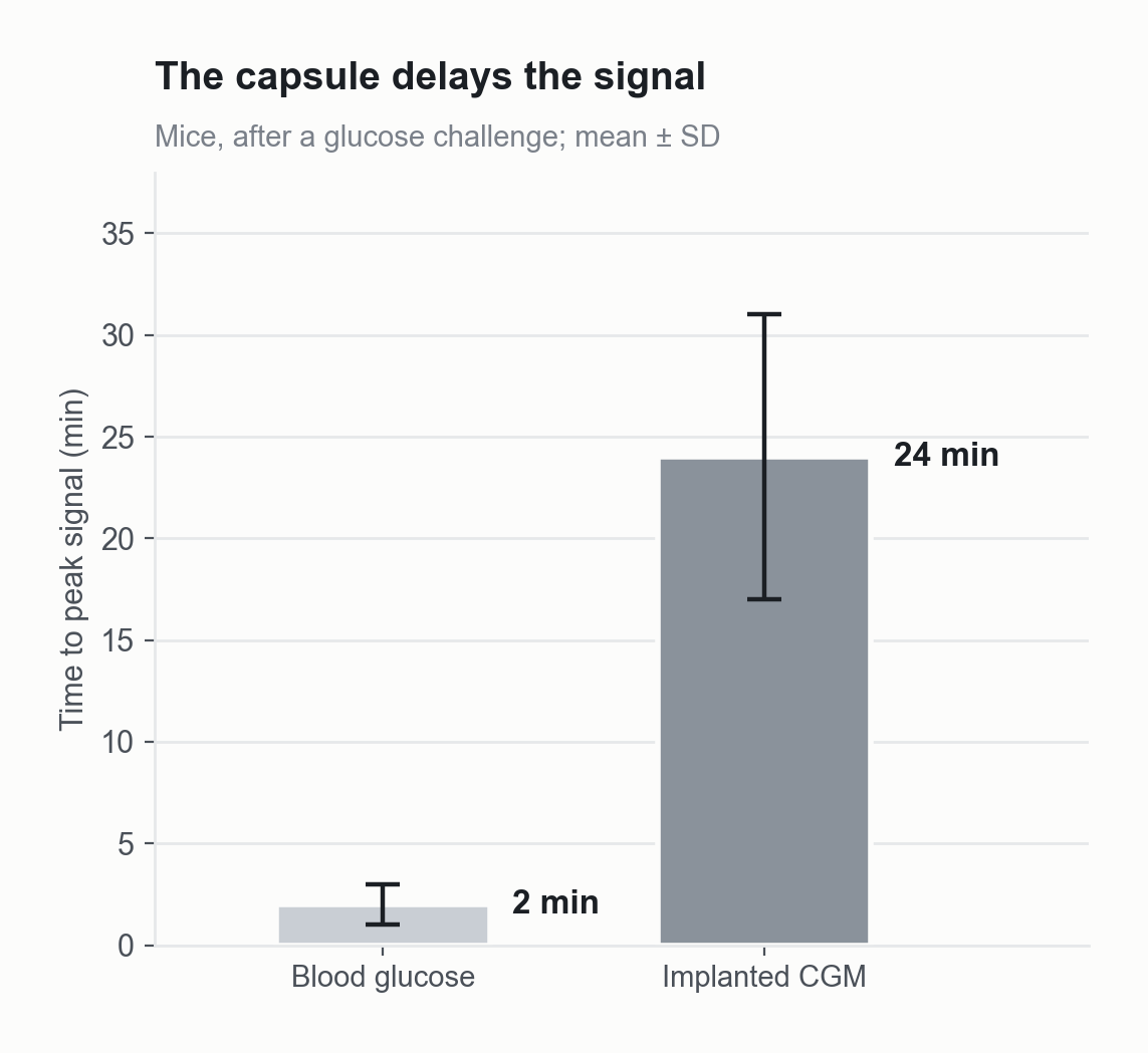
In mice, an implanted continuous glucose monitor took 24 minutes to reach its peak signal, compared with 2 minutes for blood glucose.
Source: McClatchey PM, McClain ES, Williams IM, et al. Fibrotic encapsulation is the dominant source of continuous glucose monitor delays. Diabetes 2019;68(10):1892-1901. doi:10.2337/db19-0229
The problem
The body’s response to a sensor limits its accuracy and its wear life. In mice, an implanted continuous glucose monitor took 24 minutes to reach its peak signal, against 2 minutes for blood glucose. Over 80 percent of that delay came from the fibrous capsule around the sensor (McClatchey 2019). The chart above shows it. The capsule shows up as lag, drift and lost sensitivity. One implantable sensor controls the tissue response with a steroid-eluting collar. That is a finite drug reservoir.
What ZwiGuard does here
No sensor has been coated and measured in a published ZwiGuard study. The results below are on silicone and on implant arrays.
Scar capsule, in mice. Around coated silicone sheets at six weeks the capsule was 27 micrometres, against 82 uncoated (p = 0.010). Around human cochlear implant arrays at one year it was about 60 percent thinner, 47 against 18 micrometres, including over platinum contacts (p = 0.026). The film was intact at one year. (Horne 2023.)
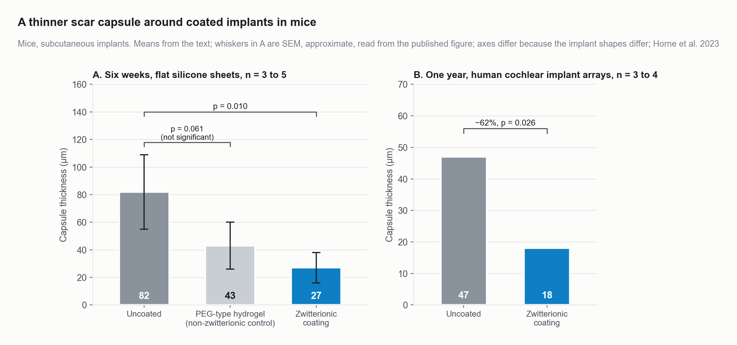
In mice, scar capsules were 67 percent thinner around zwitterionic-coated silicone at six weeks and 62 percent thinner around coated human cochlear implant arrays at one year; a non-zwitterionic hydrogel control did not differ significantly from uncoated.
Source: Horne R, Ben-Shlomo N, Jensen M, et al. Reducing the foreign body response on human cochlear implants and their materials in vivo with photografted zwitterionic hydrogel coatings. Acta Biomaterialia 2023;166:212-223, Figs 5D and 7I (means and p values from the text; SEM whiskers approximate, read from the published figure). doi:10.1016/j.actbio.2023.05.011
Cells and fibrosis, in sheep. At the round window of the sheep cochlea, coated arrays had fewer cell nuclei, fewer macrophages and less fibrosis than uncoated arrays when the three sheep were pooled. One sheep showed no difference in cells or macrophages. (Ben-Shlomo 2026.)
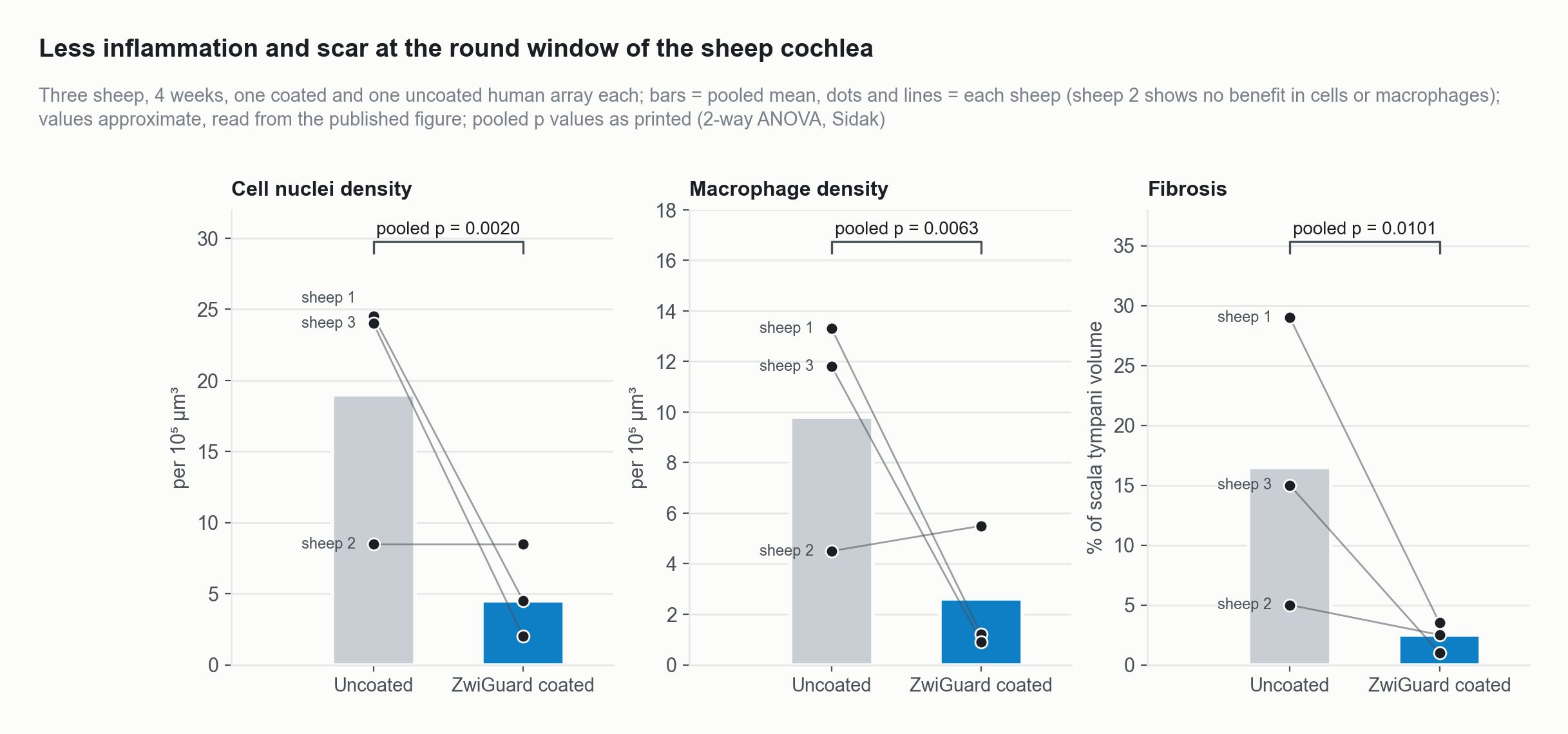
At the round window of the sheep cochlea, coated arrays had fewer cell nuclei, fewer macrophages and less fibrosis than uncoated arrays when the three sheep were pooled. One sheep showed no difference in cells or macrophages.
Source: Ben-Shlomo N, et al. Hearing Research 2026;474:109606, Fig 6E to 6G (bar and per-sheep values approximate, read from the published figure; pooled p values as printed). doi:10.1016/j.heares.2026.109606
Precedent for the class. A different zwitterionic coating, poly(MPC), cut glucose sensor error without recalibration from 73 to 17 percent in mice over three days and from 48 to 18 percent in non-diabetic primates over five days (Xie 2018). In diabetic primates the gap was smaller, 32 against 22 percent, and it disappeared with recalibration. That is not a ZwiGuard result. ZwiGuard is a thicker, crosslinked, grafted hydrogel rather than a brush.
Why it drops in
One coating step goes onto the sensor body. The published silicone films were about 7 micrometres thick (Leigh 2019). A sensor needs the film to stay thin and let glucose through, and the film can be tuned. More crosslinker makes the gel stiffer and less swollen, while the protein, cell and friction benefits are strongest at about 5 to 15 percent crosslinker.
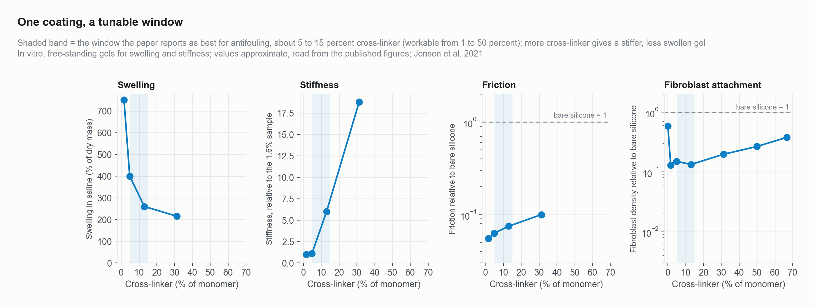
For the coating, more cross-linker makes the gel stiffer and less swollen, while protein, cell and friction benefits are strongest at about 5 to 15 percent cross-linker.
Source: Jensen MJ, Peel A, Horne R, Chamberlain J, Xu L, Hansen MR, Guymon CA. ACS Biomaterials Science and Engineering 2021;7(9):4494-4502, Figs 3G, 4A, 4B, 5B (SBMA series; values approximate, read from the published figures; stiffness shown relative to the lowest cross-linker, baseline implied by the text, 50-fold rise; the 5 to 15 percent window is stated in the text). doi:10.1021/acsbiomaterials.1c00852
Glucose diffusion through the film and sensor response time are not yet measured. Your bench measures response time on the coated parts. The film is drug-free.
How to start
Send sensor bodies, membrane coupons and a few working sensors; we return them coated, with matched controls and our adhesion data, and you measure the response. Start a feasibility round
References (6)
- McClatchey PM, McClain ES, Williams IM, et al. Fibrotic encapsulation is the dominant source of continuous glucose monitor delays. Diabetes. 2019;68(10):1892-1901.
- Xie X, et al. Zwitterionic pMPC coating of continuous glucose monitor sensors. Nat Biomed Eng. 2018 (as cited in the ZwiCoat sensors deck).
- Horne R, Ben-Shlomo N, Jensen M, et al. Reducing the foreign body response on human cochlear implants and their materials in vivo with photografted zwitterionic hydrogel coatings. Acta Biomater. 2023;166:212-223.
- Peel A, Bennion D, Horne R, Hansen MR, Guymon CA. Photografted zwitterionic hydrogel coating durability for reduced foreign body response to cochlear implants. ACS Appl Bio Mater. 2024;7(5):3124-3135.
- Ben-Shlomo N, Peel A, Etler C, et al. Development of zwitterionic hydrogel thin film coatings of cochlear implant electrode arrays that reduce intracochlear inflammation and electrode impedance. Hear Res. 2026;474:109606.
- Leigh BL, Cheng E, Xu L, Derk A, Hansen MR, Guymon CA. Antifouling photograftable zwitterionic coatings on PDMS substrates. Langmuir. 2019;35(4):1100-1110.