
Application
Neural interfaces
ZwiGuard™ is a drug-free coating that keeps scar and protein off the electrode surface, tested on human cochlear implant arrays in mice, sheep and cadaveric cochleae.
- Devices
- Cochlear implant arrays, deep brain and spinal cord leads, cortical arrays
- Substrates
- silicone and platinum contacts (published); PEEK, parylene-C and polyurethane (ZwiCoat internal tests); others in the feasibility round
- Thickness
- tunable, from a few micrometres to 100 micrometres or more (ZwiCoat process range); about 25 micrometres hydrated on sheep arrays (Ben-Shlomo 2026); about 7 micrometres in the published silicone work (Leigh 2019)
- Sterilization
- validated on your cycle during joint development
- Regulatory path
- PMA supplement for approved neurostimulators; our Master File supports the filing
- Evidence
- mice, one year; sheep, four weeks; human cadaveric cochleae
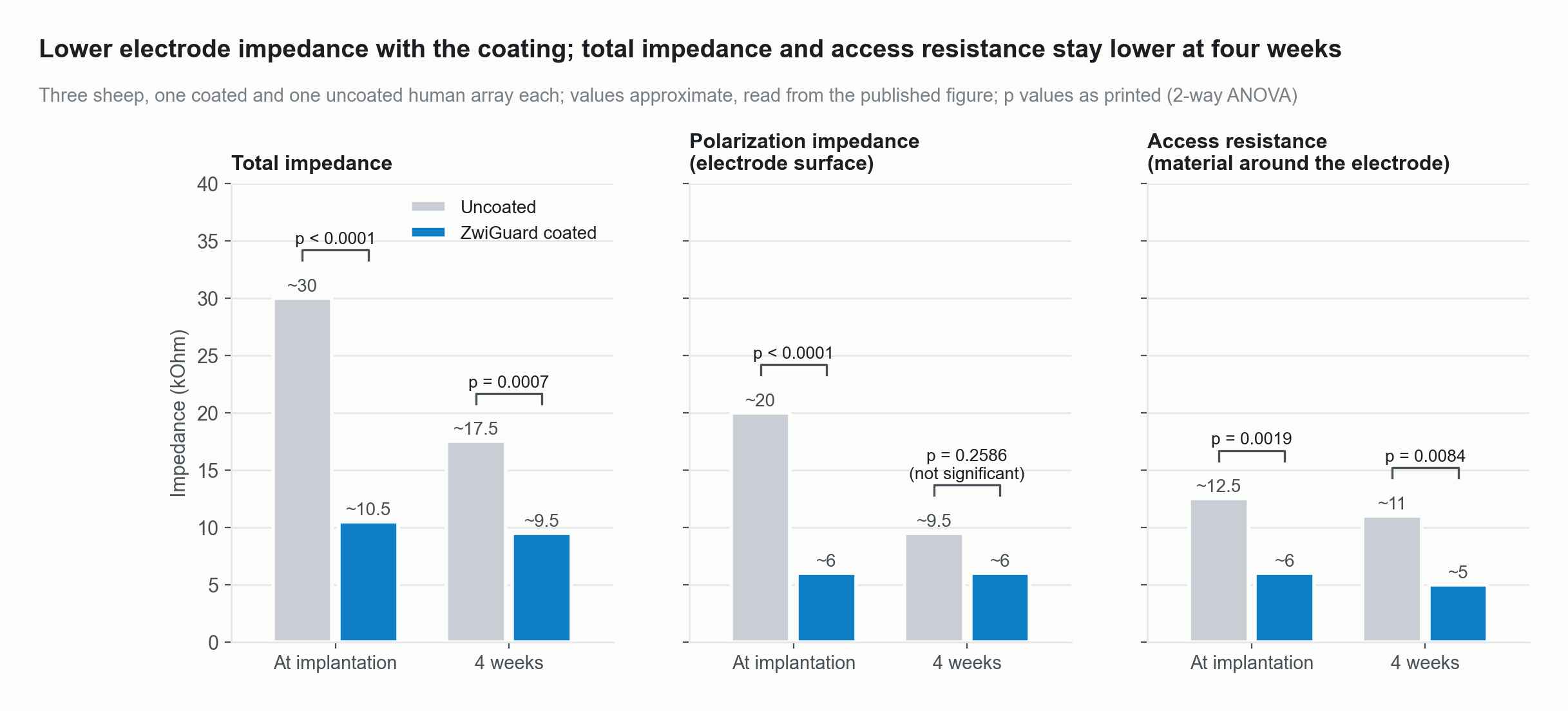
Total impedance, polarization impedance and access resistance of coated and uncoated human cochlear implant arrays in sheep, at implantation and at four weeks.
Source: Ben-Shlomo N, et al. Hearing Research 2026;474:109606, Fig 3B to 3D (bar heights approximate, read from the published figure; p values as printed). doi:10.1016/j.heares.2026.109606
The problem
A neural electrode works or fails at one place, the interface between the device and tissue. Within seconds of implantation, proteins coat the array. Over weeks, scar tissue walls it off. That scar sits in the electrical path. It can raise impedance, so stimulation needs more voltage and power. It can spread current, so channels lose selectivity. Better geometry, softer substrates and steroid elution help at the margins. None of them changes the surface where the response starts.
What ZwiGuard does here
Every result below comes from a published study. No coated device has been implanted in a person.
Impedance, in sheep. The chart above shows it. Three sheep each received one coated and one uncoated human cochlear implant array. Total impedance was about 30 against 10.5 kilohms at implantation (p < 0.0001) and about 17.5 against 9.5 at four weeks (p = 0.0007), about 50 percent lower. Access resistance was also lower at four weeks (p = 0.0084). Polarization impedance was not significantly different then (p = 0.26). (Ben-Shlomo 2026, Hearing Research.)
Inflammation, in sheep. At the round window, the pooled coated ears had fewer cell nuclei (p = 0.0020), fewer macrophages (p = 0.0063) and less fibrous tissue (p = 0.0101). One of the three sheep showed no difference in cells or macrophages. At the basal turn the pooled differences were not significant. The authors call the study a small pilot. (Ben-Shlomo 2026.)
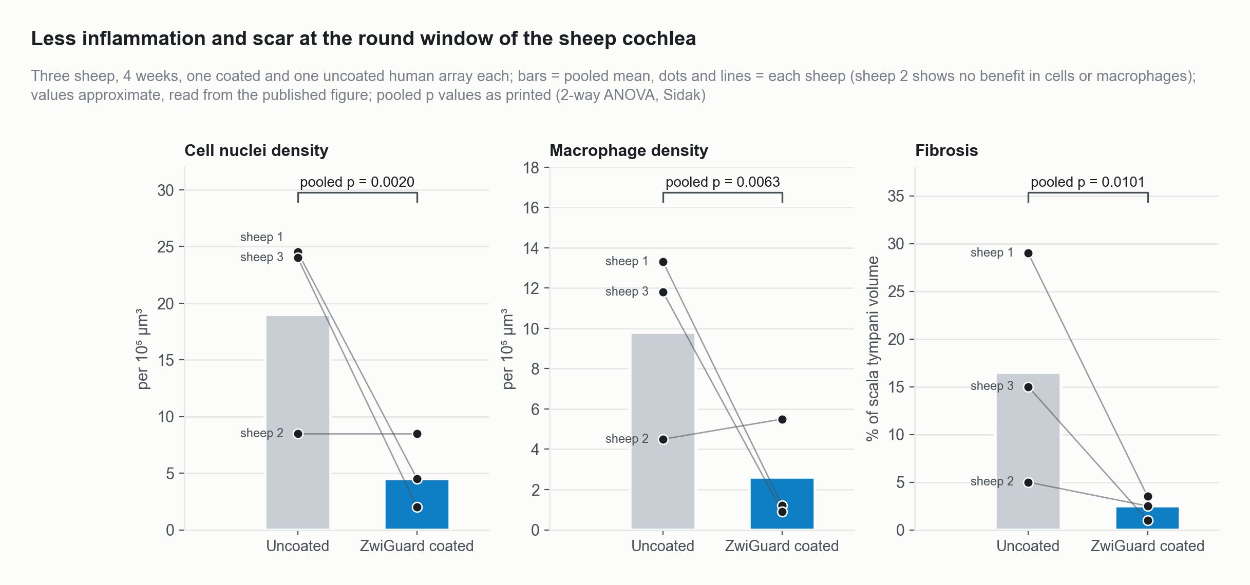
At the round window of the sheep cochlea, coated arrays had fewer cell nuclei, fewer macrophages and less fibrosis than uncoated arrays when the three sheep were pooled. One sheep showed no difference in cells or macrophages.
Source: Ben-Shlomo N, et al. Hearing Research 2026;474:109606, Fig 6E to 6G (bar and per-sheep values approximate, read from the published figure; pooled p values as printed). doi:10.1016/j.heares.2026.109606
Scar capsule, in mice. Human cochlear implant arrays implanted under the skin for one year grew a capsule about 60 percent thinner when coated, 47 against 18 micrometres (p = 0.026). The thin capsule held over platinum contacts as well as silicone. (Horne 2023, Acta Biomaterialia.)
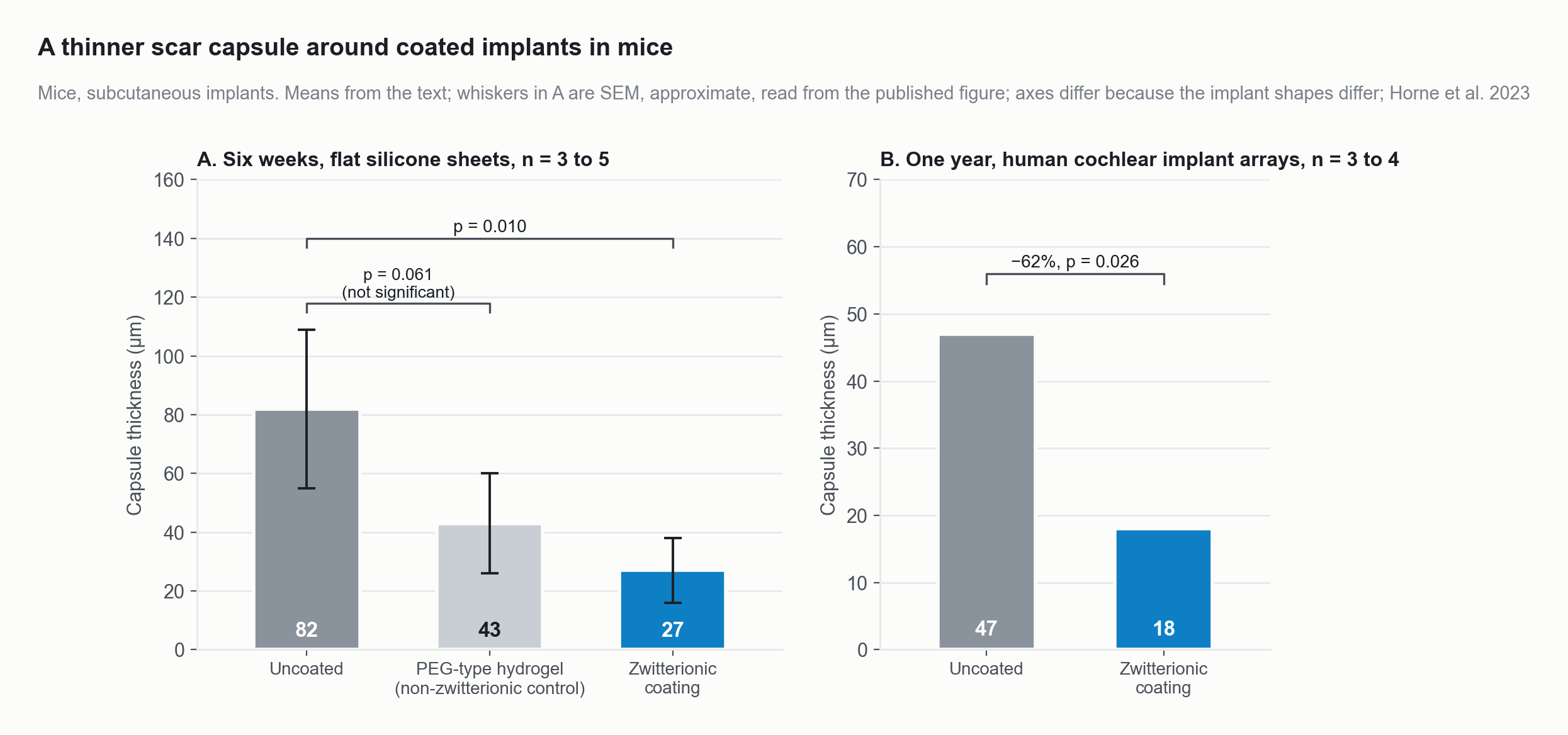
In mice, scar capsules were 67 percent thinner around zwitterionic-coated silicone at six weeks and 62 percent thinner around coated human cochlear implant arrays at one year; a non-zwitterionic hydrogel control did not differ significantly from uncoated.
Source: Horne R, Ben-Shlomo N, Jensen M, et al. Reducing the foreign body response on human cochlear implants and their materials in vivo with photografted zwitterionic hydrogel coatings. Acta Biomaterialia 2023;166:212-223, Figs 5D and 7I (means and p values from the text; SEM whiskers approximate, read from the published figure). doi:10.1016/j.actbio.2023.05.011
Insertion force, in human cadaveric cochleae. Coated arrays needed about half the peak force. In two studies it fell about 42 percent, 64.2 against 37.0 millinewtons (Bennion 2021, Otology and Neurotology; p = 0.001, nine insertions per group), and about 50 percent, about 68 against 35 millinewtons (Peel 2024, ACS Applied Bio Materials; read from the figure). Force variation fell about 42 percent in the first study.
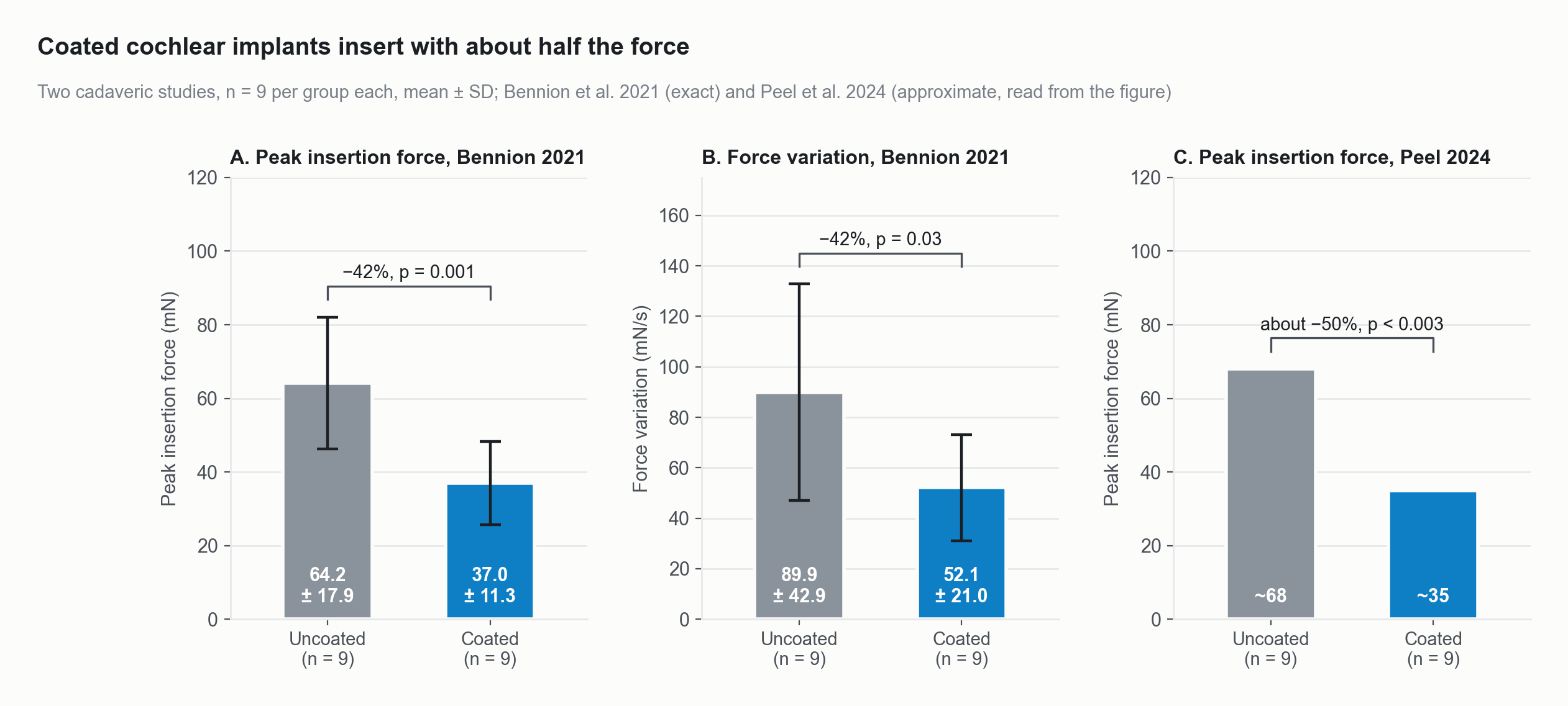
Coated cochlear implant arrays needed about half the peak insertion force of uncoated arrays in two human cadaveric studies: about 42 percent less (Bennion 2021) and about 50 percent less (Peel 2024).
Source: Bennion DM, Horne R, Peel A, Reineke P, Henslee A, Kaufmann C, Guymon CA, Hansen MR. Otology and Neurotology 2021;42(10):1476-1483, Fig 4A and 4B (means and SD from the text), doi:10.1097/MAO.0000000000003288; Peel A, Bennion D, Horne R, Hansen MR, Guymon CA. ACS Applied Bio Materials 2024;7:3124-3135, Fig 13A (values approximate, read from the published figure), doi:10.1021/acsabm.4c00156.
Under the microscope
A cross-section of a coated array, mapped by element, shows the hydrogel film on the silicone carrier. The second picture is a coated array dyed blue so the film is easy to see.
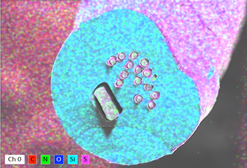
Cross-section of a coated cochlear implant array: elemental map showing the hydrogel film on the silicone carrier. ZwiCoat.
ZwiCoat
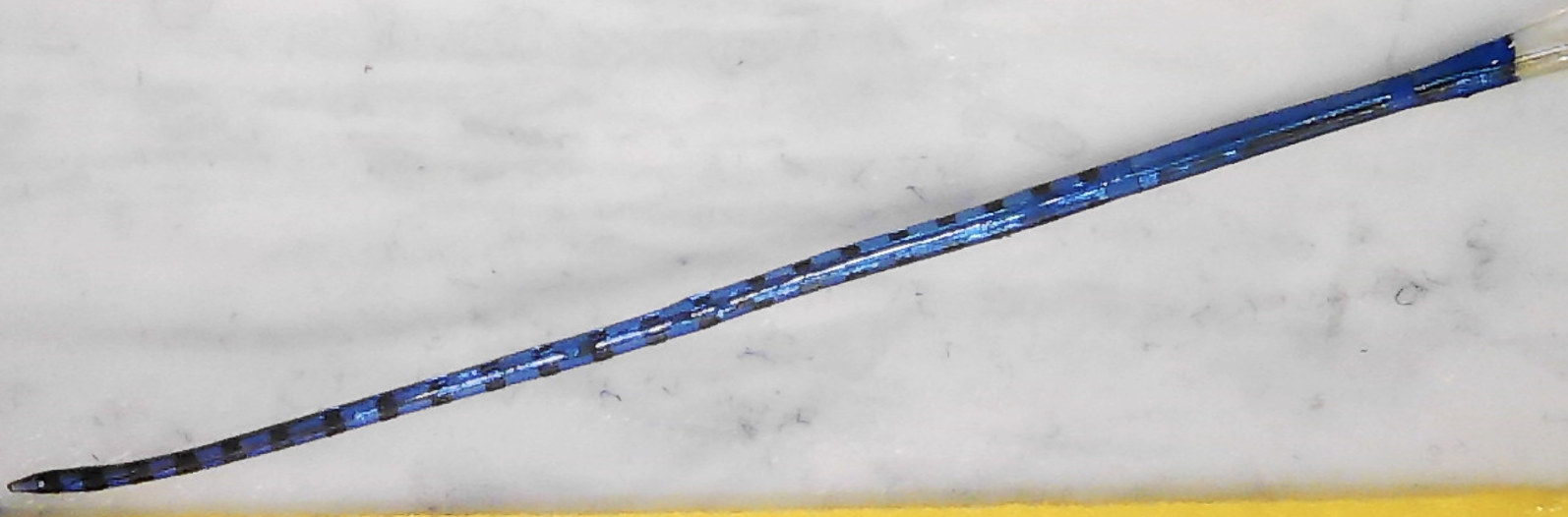
A cochlear implant array coated with the blue-dyed hydrogel.
ZwiCoat, 2026
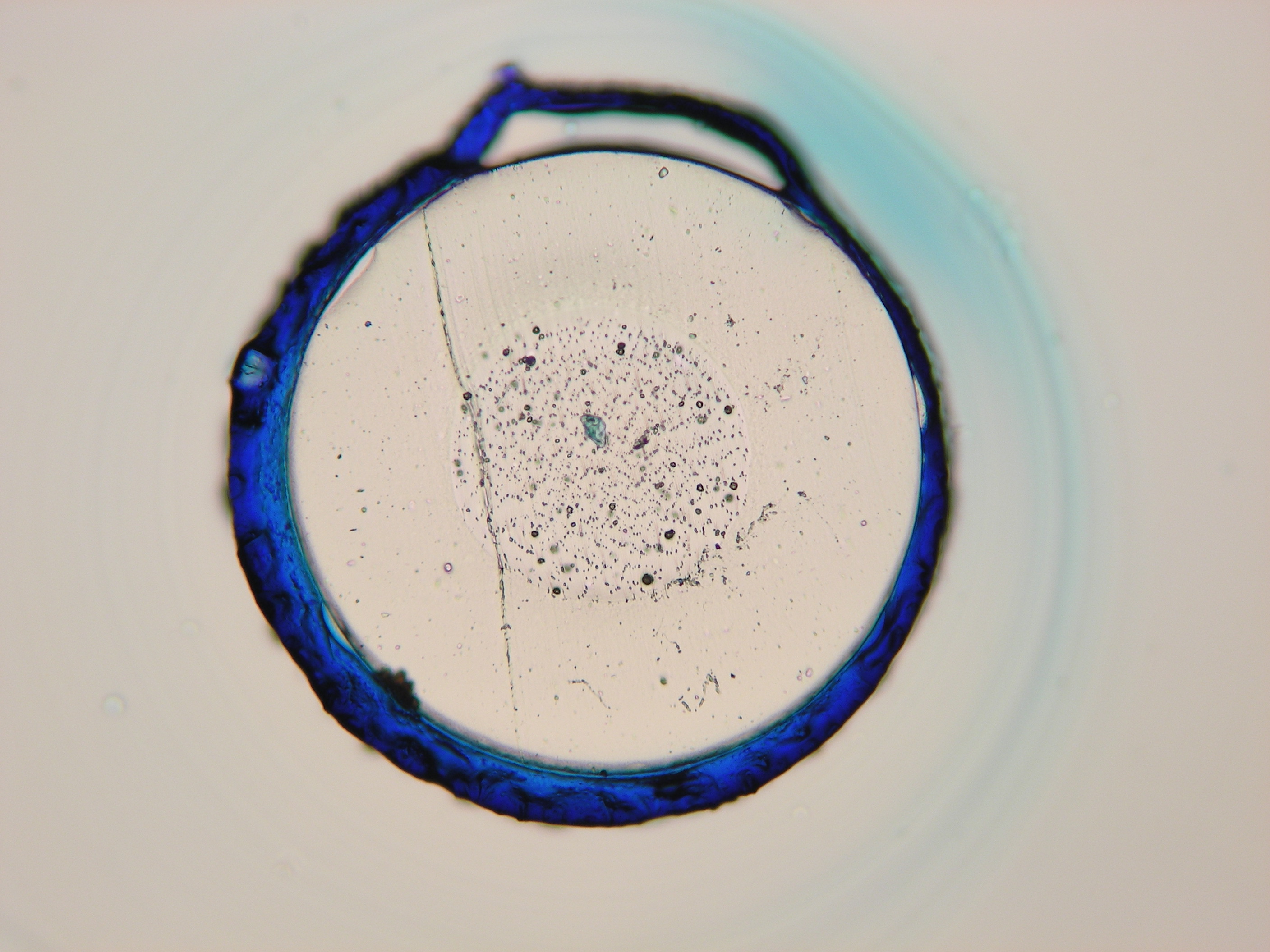
Cross-section under the microscope: a silicone cochlear implant proxy (0.635 mm core) with the dyed hydrogel coating as the blue ring around it. ZwiCoat, 2025.
ZwiCoat, 2026
In sheep, tissue sections at the round window show the difference the counts describe. The uncoated array sits in dense tissue, and the coated one in a largely clear space.
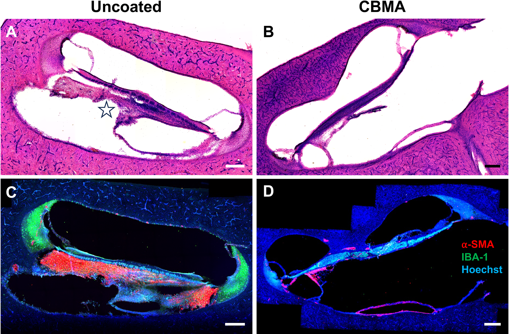
Sheep cochlea at the round window, uncoated array on the left and coated array on the right. Top row: H&E stain. Bottom row: stains for fibrosis (alpha-SMA, red), macrophages (Iba1, green) and nuclei (Hoechst, blue).
Ben-Shlomo et al. 2026, Hearing Research 474:109606, Fig 6A to D, CC BY 4.0.
Why it drops in
One UV step grafts the hydrogel to the surface and crosslinks it. The sheep arrays were coated in a printed mold, giving a film about 25 micrometres thick when hydrated, over the silicone carrier and the platinum contacts. On silicone, adhesion tests show the film failing inside the gel before the interface lets go (Leigh 2019). Bonding to your lead material is measured in the feasibility round.
How to start
Send leads or arrays and we coat them at no cost, then return them with matched uncoated controls and our data on coverage, adhesion and friction. Start a feasibility round
References (4)
- Ben-Shlomo N, Peel A, Etler C, et al. Development of zwitterionic hydrogel thin film coatings of cochlear implant electrode arrays that reduce intracochlear inflammation and electrode impedance. Hear Res. 2026;474:109606.
- Horne R, Ben-Shlomo N, Jensen M, et al. Reducing the foreign body response on human cochlear implants and their materials in vivo with photografted zwitterionic hydrogel coatings. Acta Biomater. 2023;166:212-223.
- Bennion DM, Horne R, Peel A, et al. Zwitterionic photografted coatings of cochlear implant biomaterials reduce friction and insertion forces. Otol Neurotol. 2021;42(10):1476-1483.
- Leigh BL, Cheng E, Xu L, Derk A, Hansen MR, Guymon CA. Antifouling photograftable zwitterionic coatings on PDMS substrates. Langmuir. 2019;35(4):1100-1110.